 |
ULTRASOUND
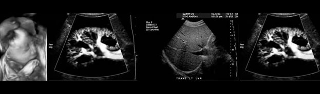
What is Ultrasound Imaging? Ultrasound imaging, also called ultrasound scanning or sonography, is a method of obtaining images from inside the human body through the use of high-frequency sound waves. The reflected sound wave echoes are recorded and displayed as a real-time visual image. No ionizing radiation (x-ray) is involved in ultrasound imaging. Obstetric ultrasound refers to the specialized use of sound waves to visualize and thus determine the condition of a pregnant woman and her embryo or fetus. Ultrasound is a useful way of examining many of the body's internal organs, including but not limited to the heart, liver, gallbladder, spleen, pancreas, kidneys and bladder. Because ultrasound images are captured in real time, they can show movement of internal tissues and organs and enable physicians to see blood flow and heart valve functions. This can help to diagnose a variety of heart conditions and to assess damage after a heart attack or other illness. What are some common uses of the procedure? Millions of expectant parents have seen the first "picture" of their unborn child with pelvic ultrasound examinations of the uterus and fetus. Ultrasound imaging is used extensively for evaluating the eyes, pelvic and abdominal organs, heart and blood vessels, and can help a physician determine the source of pain, swelling or infection in many parts of the body. Because ultrasound provides real-time images it can also be used to guide procedures such as needle biopsies, in which needles are used to sample cells from organs for laboratory testing. Ultrasound is now being used to image the breasts and to guide biopsy of breast cancer (see the Ultrasound-Guided Breast Biopsy page). Ultrasound is also used to evaluate superficial structures such as the thyroid gland and scrotum (testicles). Doppler ultrasound is a special technique used to examine blood flow. Doppler images can help the physician to see and evaluate:
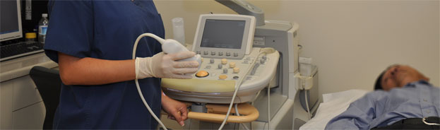
How should I prepare for the procedure? You should wear comfortable, loose-fitting clothing for your ultrasound exam. Other preparation depends on the type of examination you will have. For some scans your doctor may instruct you not to eat or drink for as many as 12 hours before your appointment. For others you may be asked to drink up to six glasses of water two hours prior to your exam and avoid urinating so that your bladder is full when the scan begins. How is the procedure performed? The patient is usually positioned on an examination table. A clear gel is applied to the patient's body in the area to be examined to help the transducer make secure contact with the skin. The sound waves produced by the transducer cannot penetrate air so the gel helps eliminate air pockets between the transducer and the skin. The technologist or radiologist presses the transducer firmly against the skin and sweeps it back and forth to image the area of interest. When the examination is complete the patient may be asked to dress and wait while the ultrasound images are reviewed either on film or on a TV monitor. Often though, the technologist or radiologist is able to review the ultrasound images in real time as they are acquired and the patient can be released immediately. What are the benefits vs. risks? Benefits
Risks
What are the limitations of General Ultrasound Imaging? Ultrasound has difficulty penetrating bone and therefore can only see the outer surface of bony structures and not what lies within. For visualization of bone, other imaging modalities such as magnetic resonance imaging (MRI) may be selected. Ultrasound waves do not pass through air; therefore an evaluation of the stomach, small intestine and large intestine may be limited. Intestinal gas may also prevent visualization of deeper structures such as the pancreas and aorta. Patients suffering from obesity are more difficult to image-this is because tissue attenuates (weakens) the sound waves as they pass deeper into the body. Source: radiologyinfo.org |
|
|