What is MRI
Magnetic resonance imaging (MRI) uses radiofrequency waves and a strong magnetic field rather than x-rays to provide remarkably clear and detailed pictures of internal organs and tissues. The technique has proven very valuable for the diagnosis of a broad range of conditions in all parts of the body including cancer, heart and vascular disease, stroke, joint and musculoskeletal disorders. MRI requires specialized equipment and expertise and allows evaluation of some body structures that may not be as visible with other imaging methods.
What are some common uses of the MRI procedure?
Brain:
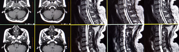
The MRI procedure has been proven to be very helpful to radiologists in diagnosing tumors of the brain as well as disorders of the eyes and the inner ear. MRI is also the most sensitive exam for strokes and certain chronic disorders of the nervous system such as multiple sclerosis. In addition, it is a useful means of documenting brain abnormalities in patients with dementia and it is commonly used for patients with disease of the pituitary gland. It requires specialized equipment and expertise and allows evaluation of some body structures that may not be as visible with other imaging methods.
Body:
Because MRI can give such clear pictures of soft-tissue structures near and around bones, it is the most sensitive exam for spinal and joint problems. MRI is widely used to diagnose sports-related injuries, especially those affecting the knee, shoulder, hip, elbow and wrist. The images allow the physician to see even very small tears and injuries to ligaments and muscles.
In addition, MRI of the heart, aorta, and blood vessels is a fast, noninvasive tool for diagnosing. Physicians can examine the size and thickness of the chambers of the heart and determine the extent of damage caused by a heart attack or progressive heart disease.
Organs of the chest and abdomen-including the lungs, liver , kidney , spleen , pancreas and abdominal vessels-can also be examined in high detail with MRI, enabling the diagnosis and evaluation of tumors and functional disorders. MRI is growing in popularity as an alternative to traditional x-ray. MRI can supplement mammography in the early diagnosis of breast cancer. Because no radiation exposure is involved, MRI is often the preferred diagnostic tool for the examination of the male and female reproductive systems, pelvis and hips and the bladder.
How should I prepare for the procedure?
Because the strong magnetic field used for MRI will pull on any ferromagnetic metal object implanted in the body, MRI staff will ask whether you have a prosthetic hip, heart pacemaker (or artificial heart valve), implanted port , infusion catheter (brand names Port-o-cath , Infusaport , Lifeport ), intrauterine device (IUD), or any metal plates, pins, screws or surgical staples in your body. In most cases surgical staples, plates, pins and screws pose no risk during MRI if they have been in place for more than four to six weeks. Tattoos and permanent eyeliner may also create a problem. You will be asked if you have ever had a bullet or shrapnel in your body or ever worked with metal. If there is any question of metal fragments, you may be asked to have an x-ray that will detect any such metal objects. Tooth fillings usually are not affected by the magnetic field but they may distort images of the facial area or brain, so the radiologist should be aware of them. The same is true of braces, which may make it hard to "tune" the MRI unit to your body. You will be asked to remove anything that might degrade MRI images of the head, including hairpins, jewelry, eyeglasses, hearing aids and any removable dental work.
The radiologist or technologist may ask about drug allergies and whether head surgery has been done in the past. If you might be pregnant, this should be mentioned. Some patients who undergo MRI in an enclosed unit may feel confined or claustrophobic. If you are not easily reassured, a sedative may be administered. Roughly one in 20 patients will require medication to reduce the anxiety associated with claustrophobia.
How is the procedure performed?
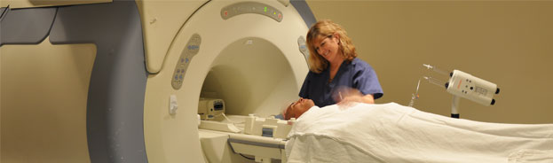
The patient is placed on a sliding table and positioned comfortably for the MRI examination. Then the radiologist and technologist leave the room and the individual MRI sequences are performed. The patient is able to communicate with the radiologist or technologist at any time using an intercom. Also, many MRI centers allow a friend or, if a child is being examined, a parent to stay in the room. Depending on how many images are needed, the exam will generally take 15 to 45 minutes, although a very detailed study may take longer. You will be asked not to move during the actual imaging process, but between sequences some movement is allowed. Patients are generally required to remain still for only a few seconds to a few minutes at a time.
Depending on the part of the body being examined, a contrast material (usually gadolinium) may be used to enhance the visibility of certain tissues or blood vessels. A small needle connected to an intravenous line is placed in an arm or hand vein. A saline solution will drip through the intravenous line to prevent clotting until the contrast material is injected about two-thirds of the way through the exam.
When the exam is over the patient is asked to wait until the images are examined to determine if more images are needed. A radiologist experienced in MRI will analyze the images and send a report with his or her interpretation to the patient's personal physician. This should take only a few days or less.
What are the benefits vs. risks?
Benefits
- Images of the soft-tissue structures of the body-such as the brain, spine, joints, liver and other organs-are clearer and more detailed than with other imaging methods.
- MRI can help physicians evaluate the function as well as the structure of many organs.
- The detail makes MRI an invaluable tool in early diagnosis and evaluation of tumors.
- MRI contrast material is less likely to produce an allergic reaction than the iodine-based materials used for conventional x-rays and CT scanning.
- MRI enables the detection of abnormalities that might be obscured by bone with other imaging methods.
- MRI provides a fast, noninvasive alternative to x-ray angiography for diagnosing problems of the cardiovascular system.
- Exposure to radiation is avoided.
Risks
- An undetected metal implant may be affected by the strong magnetic field.
- MRI is generally avoided in the first 12 weeks of pregnancy. Doctors usually use other methods of imaging, such as ultrasound , on pregnant women unless there is a strong medical reason to use MRI.
MR Angiography (MRA)
What is MR Angiography?
MR angiography (MRA) is an MRI study of the blood vessel s. It utilizes MRI technology to detect, diagnose, and aid the treatment of heart disorders, stroke, and blood vessel diseases. For select MRA exams, a special form of contrast material is often given to make the MRI images even clearer. The procedure is painless, and the magnetic field is not known to cause tissue damage of any kind.
What are some common uses of the MRA procedure?
Many patients with arterial disease now have it treated in the radiology department rather than undergoing surgery in an operating room. MRA is a very useful way of finding problems with blood vessels and determining how to best treat those problems.
The carotid arteries in the neck that conduct blood to the brain are a common site of atherosclerosis, which may severely narrow or block off an artery, reducing blood flow to the brain and even causing a stroke. If an ultrasound study shows that such disease is present, many surgeons will perform the necessary operation after confirmation with MRA, dispensing with the need for catheter angiography.
MRA has found wide use in checking patients for diseased intracranial (in the head) arteries, so that only those with positive findings will need to undergo a more invasive catheter study.
MRA is also used to detect disease in the aorta and in blood vessels supplying the kidneys, lungs, and legs.
Patients with a family history of arterial aneurysm, a ballooning out of a segment of the vessel wall, can be screened with MRA to see if they have a similar disorder that has not produced symptoms. If an aneurysm is found, it may be eliminated surgically, possibly avoiding serious or fatal bleeding.
Source: RadiologyInfo